A Model for Image Segmentation under Noisy Conditions and Anomaly Detection in Medical MRI Images Using an RBF Neural Network and the PSO Metaheuristic Algorithm for Breast Cancer Detection
Keywords:
Radial Basis Function (RBF) neural network, Particle Swarm Algorithm (PSO), Metaheuristic Algorithms, Hybrid Algorithm Design, MATLAB Simulation, Intelligent SegmentationAbstract
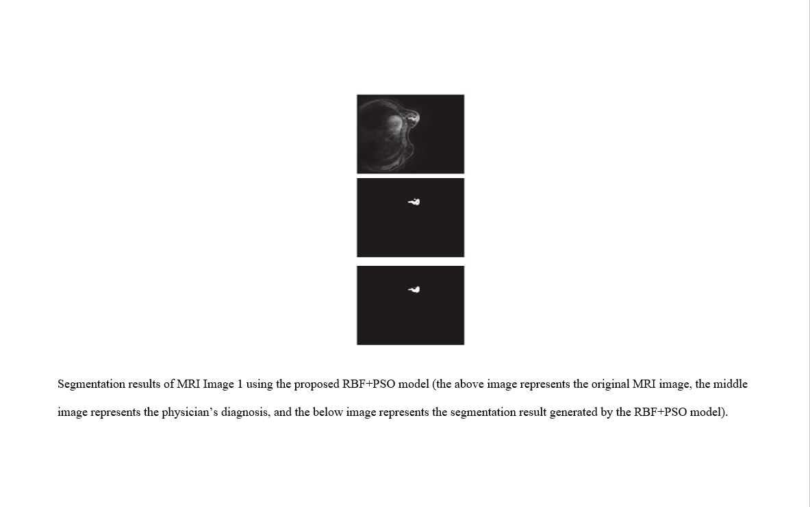
The aim of the present study was to develop an intelligent model for image segmentation under noisy conditions and anomaly detection in medical MRI images using a Radial Basis Function (RBF) neural network and the Particle Swarm Optimization (PSO) metaheuristic algorithm. In this model, the RBF neural network was responsible for learning and separating different image regions, while the PSO algorithm was employed for the automatic optimization of the network parameters, including centers, weights, and radii of the basis functions. The MRI images used in this study consisted of real data from the breast tissues of patients with and without cancer. Following the preprocessing stage, which included Gaussian and salt-and-pepper noise removal and pixel intensity normalization, the images were applied to the proposed model. The model output provided accurate segmentation of regions suspected of abnormalities from other tissues, and the results were compared with classical K-Means, FCM, and standalone RBF network methods. The findings demonstrated that the hybrid RBF–PSO model was able to increase segmentation accuracy under noisy conditions to an average of 96.4% and achieved a Dice overlap index of 0.92, representing a significant improvement compared to the other methods. Furthermore, the proposed model showed stable performance in preserving the actual tumor boundaries and eliminating noisy regions. Based on the obtained results, the RBF–PSO model can be considered an efficient tool for the early detection of breast cancer through the automated analysis of MRI images. It is recommended that future studies integrate this model with deep learning networks in order to improve its accuracy and generalizability in clinical datasets.
References
[1] F. Sadoughi, Z. Kazemy, F. Hamedan, L. Owji, M. Rahmanikatigari, and T. T. Azadboni, "Artificial Intelligence Methods for the Diagnosis of Breast Cancer by Image Processing: A Review," Breast Cancer: Targets and Therapy, vol. 10, p. 219, 2018.
[2] M. Muhammad, D. Zeebaree, A. M. A. Brifcani, J. Saeed, and D. A. Zebari, "Region of Interest Segmentation Based on Clustering Techniques for Breast Cancer Ultrasound Images: A Review," Journal of Applied Science and Technology Trends, vol. 1, no. 3, pp. 78-91, 2020.
[3] B. Ravi, V. Amgothu, K. Keshamoni, A. Vinisha, P. K. Poola, and M. O. Khan, "A Robust Noise Reduction Strategy in Magnetic Resonance Images," Annals of the Romanian Society for Cell Biology, vol. 25, no. 6, pp. 3938-3952, 2021.
[4] S. Sudharson and P. Kokil, "Computer-Aided Diagnosis System for the Classification of Multi-Class Kidney Abnormalities in the Noisy Ultrasound Images," Computer Methods and Programs in Biomedicine, vol. 205, p. 106071, 2021.
[5] L. Hua, Y. Gu, X. Gu, J. Xue, and T. Ni, "A Novel Brain MRI Image Segmentation Method Using an Improved Multi-View Fuzzy C-Means Clustering Algorithm," Frontiers in Neuroscience, vol. 15, p. 245, 2021.
[6] T. X. Pham, P. Siarry, and H. Oulhadj, "Integrating Fuzzy Entropy Clustering with an Improved PSO for MRI Brain Image Segmentation," Applied Soft Computing, vol. 65, pp. 230-242, 2018.
[7] G. N. Girish, B. Thakur, S. R. Chowdhury, A. R. Kothari, and J. Rajan, "Segmentation of Intra-Retinal Cysts from Optical Coherence Tomography Images Using a Fully Convolutional Neural Network Model," IEEE Journal of Biomedical and Health Informatics, vol. 23, no. 1, pp. 296-304, 2018.
[8] P. Kumar, P. Nagar, C. Arora, and A. Gupta, "U-SegNet: Fully Convolutional Neural Network Based Automated Brain Tissue Segmentation Tool," in 2018 25th IEEE International Conference on Image Processing (ICIP), 2018: IEEE, pp. 3503-3507.
[9] D. Dai, C. Dong, and S. Xu, "MS RED: A Novel Multi-Scale Residual Encoding and Decoding Network for Skin Lesion Segmentation," Medical Image Analysis, vol. 75, p. 102293, 2022.
[10] M. Soleimani, A. Harooni, N. Erfani, A. Rehman, T. Saba, and S. A. Bahaj, "Classification of Cancer Types Based on microRNA Expression Using a Hybrid Radial Basis Function and Particle Swarm Optimization Algorithm," Microscopy Research and Technique, vol. 87, no. 5, pp. 1052-1062, 2024, doi: 10.1002/jemt.24492.
[11] A. Bilal, A. Imran, T. I. Baig, X. Liu, E. A. Nasr, and H. Long, "Breast Cancer Diagnosis Using Support Vector Machine Optimized by Improved Quantum Inspired Grey Wolf Optimization," Scientific Reports, vol. 14, no. 1, 2024, doi: 10.1038/s41598-024-61322-w.
[12] F. Farheen, M. S. Shamil, N. Ibtehaz, and M. S. Rahman, "Revisiting Segmentation of Lung Tumors from CT Images," Computers in Biology and Medicine, vol. 144, p. 105385, 2022.
[13] M. Kannan and C. Priya, "A Research on Prediction of Bat-Borne Disease Infection Through Segmentation Using Diffusion-Weighted MR Imaging in Deep-Machine Learning Approach," Materials Today: Proceedings, 2021.
[14] M. K. Islam, C. Kaushal, and M. A. Amin, "A Secure Framework Toward IoMT-Assisted Data Collection, Modeling, and Classification for Intelligent Dermatology Healthcare Services," Contrast Media and Molecular Imaging, vol. 2022, pp. 6805460-6805460, 2022.
[15] R. M. Alzahrani et al., "Early Breast Cancer Detection via Infrared Thermography Using a CNN Enhanced With Particle Swarm Optimization," Scientific Reports, vol. 15, no. 1, 2025, doi: 10.1038/s41598-025-11218-0.
[16] H. Khodadadi and S. Nazem, "Improving Cancer Detection Through Computer-Aided Diagnosis: A Comprehensive Analysis of Nonlinear and Texture Features in Breast Thermograms," Plos One, vol. 20, no. 5, p. e0322934, 2025, doi: 10.1371/journal.pone.0322934.
[17] P. Capar, M. Rakhra, G. Cazzato, and M. S. Hossain, "A Novel Hybrid Deep Learning Approach for Skin Lesion Segmentation and Classification," Journal of Healthcare Engineering, pp. 1-21, 2022.
[18] S. Dash et al., "Brain Tumor Detection and Classification Using IFF‐FLICM Segmentation and Optimized ELM Model," Journal of Engineering, vol. 2024, no. 1, 2024, doi: 10.1155/2024/8419540.
[19] D. Selvathi, "Brain Tissues Segmentation in Magnetic Resonance Imaging for the Diagnosis of Brain Disorders Using a Convolutional Neural Network," in Handbook of Decision Support Systems for Neurological Disorders: Academic Press, 2021, pp. 167-186.
[20] N. V. Shree and T. N. R. Kumar, "Identification and Classification of Brain Tumor MRI Images with Feature Extraction Using DWT and Probabilistic Neural Network," Brain Informatics, vol. 5, no. 1, pp. 23-30, 2018.
Downloads
Publication Timeline
- Submitted
- Revised
- Accepted
Issue
Section
License
Copyright (c) 2025 Ehab Maan Turki AL Khafaji (Author); Hussein Moradi (Corresponding author); Hadab Khalid Obayes, Mehdi Nasri, Reihaneh Khorsand (Author)

This work is licensed under a Creative Commons Attribution-NonCommercial 4.0 International License.